As Marshall B. Ketchum University evolves into an interprofessional institution, it is preparing both its curriculum and technology for the future of medical education, in which technology is especially important to learning and growth for both students and the university.
MBKU recently purchased new equipment for its current programs representing the latest advances in medical technology: two Anatomage Tables and direct and indirect ophthalmoscopy simulators. MBKU’s PA and optometry programs will be able to use the technology together in developing curricula interprofessionally.
Accurate, comprehensive and hands-on Anatomage Tables teach virtual dissection and anatomy on large touch screens — much like a giant tablet — with life-size 3-D and 4-D (such as to show respiration) scans of actual adult male and female cadavers. The tables can be tilted vertically or horizontally for ease of use, and come with a digital library of scans representing more than 100 real-life pathological cases that show what different conditions or anatomical variations look like in different individuals, including bone fractures, aneurysms, heart conditions and rare conditions.
Students can view and dissect organs and tissue from different angles, then restore the scans to their original state and try again. Students will also be able to upload patients’ imaging scans and use the tables’ built-in software to create 3-D renderings.
The tables provide many advantages over cadavers, which MBKU’s PA program has not yet used in its short history: while the up-front cost is significant, the tables are a one-time purchase and eliminate expenses and hassles associated with properly storing and using cadavers.
MBKU is developing interprofessional courses in which students from all three of its disciplines will work together on specific case studies using the tables during class. In her neuroscience courses, Rima Khankan, PhD, assistant professor of neurosciences, plans to use the tables to demonstrate how the spinal pathways connect to the brain and to zoom in on detailed views of neurophysiological structures — especially helpful to optometry students. Meanwhile, future pharmacy students will use the equipment for virtual dissection, as PA students do, during their required year of anatomy courses.
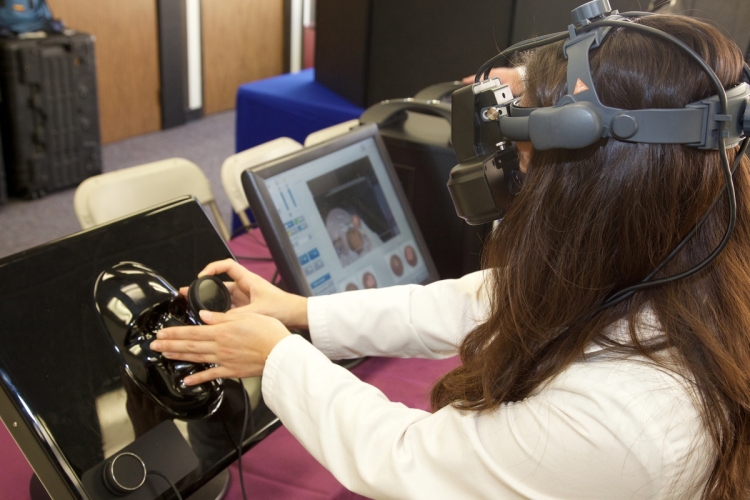
While medical education often relies on different types of simulators, not many optometric simulators exist. MBKU is among the early adopters of the Eyesi Indirect and Eyesi Direct Ophthalmoscope Simulators, which perform as their names suggest by having students look through the devices to practice ophthalmoscopy exams on virtual patients rather than on classmates or significant others. The simulators also provide consistency in learning, as no two students who practice solely on human subjects will receive the same experience.
The simulators give students a broader view of the retina — a scope that is often limited in human patients — and gives direct procedural and diagnostic feedback to fine-tune students’ skills before they perform real-life examinations. To use the indirect simulator, students use a head-worn device and hold a lens up to a 3-D model attached to a computer; the direct simulator contains a handheld lens, a freestanding mannequin head and a touch-screen computer. Like the Anatomage Table, the simulators are loaded with case studies and pathology examples from real patients. This case-based learning method gives students confidence and competence in diagnostic capabilities and critical thinking.
Associate Dean of Academic Affairs Raymond Chu, OD, MS, plans to incorporate the simulators into first- and second-year optometry classrooms this spring. For PA students, ophthalmoscopy is one of many skills learned that isn’t always prioritized in the classroom. Using the simulators and receiving feedback from optometry students will give PA students a solid foundation in ophthalmoscopy.
Special thanks to Dr. Joseph and Mrs. Peggy Taylor for their generosity which made the ophthalmoscopy simulation lab possible.

